Cardiovascular System
Read/Download our Books for Free:
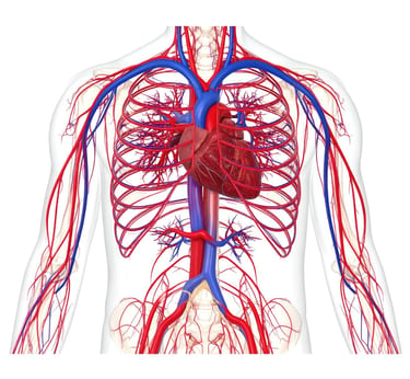
Cardiovascular System Detailed Breakdown:
1. Heart
Structure:
Four chambers: Left/Right Atria (receive blood) and Left/Right Ventricles (pump blood) [B-3].
Valves: Mitral (bicuspid), Tricuspid, Aortic, Pulmonary (semilunar) – prevent backflow [B-3].
Coronary arteries (right and left) supply oxygenated blood to the myocardium; veins drain into the coronary sinus → right atrium [B-2], [S-4].
Electrical conduction: SA node → AV node → Bundle of His → Purkinje fibers [B-3].
Function:
Dual pump: Systemic circulation (left heart) and Pulmonary circulation (right heart) [B-5].
Starling’s Law: Force of contraction depends on ventricular filling volume [B-3].
2. Arteries
Aorta:
Ascending aorta: Branches into coronary arteries [B-4].
Aortic arch: Contains baroreceptors/chemoreceptors for BP/pH regulation [B-4].
Descending aorta: Supplies thorax/abdomen [B-4].
Coronary arteries:
Left coronary artery: Divides into anterior interventricular and circumflex branches [B-4].
Right coronary artery: Supplies SA/AV nodes; ends as posterior interventricular artery [B-4].
Unique in elasmobranchs: Terminate in Thebesian vessels draining into ventricular lumen [S-4].
Pathology:
Atherosclerosis: Plaque buildup (LDL-driven) narrows arteries; garlic extract shown to decalcify plaques [A-15], [S-6].
Coarctation: Congenital aortic narrowing → hypertension (treated via angioplasty/stenting) [A-9].
3. Veins
Superior/Inferior Vena Cava: Return deoxygenated blood to right atrium [B-1].
Pulmonary veins (4 total): Carry oxygenated blood from lungs to left atrium [B-1].
Portal circulation: Nutrient-rich blood from GI tract → liver via hepatic portal vein [B-5].
Varicose veins: Caused by valve incompetence; exacerbated by low-fiber diets [B-5].
4. Capillaries
Structure: Thin-walled (single endothelial layer) for gas/nutrient exchange [B-2].
Types:
Continuous: Least permeable (e.g., blood-brain barrier).
Fenestrated: Pores for filtration (e.g., kidneys) [B-2].
Function:
Hydrostatic pressure forces fluid out at arterial end; osmotic pressure draws it back at venous end [B-2].
Shunts in tadpole gills divert blood flow during metamorphosis [S-2].
5. Blood
Components:
Plasma (55%): Water, proteins (albumin, globulins), electrolytes.
Cells (45%): Erythrocytes (O₂/CO₂ transport), Leukocytes (immunity), Platelets (clotting) [B-5].
Pathology:
Hypercholesterolemia: Poor correlation with atherosclerosis in autopsy studies [A-4].
Garlic’s role: Reduces LDL, triglycerides; inhibits platelet aggregation [A-8], [A-1].
6. Key Natural Therapies (from Articles)
Garlic: Lowers BP, reduces arterial stiffness, and decalcifies plaques [A-1], [A-15].
Vitamin C + Lysine: Linus Pauling Protocol reverses plaque by strengthening collagen [A-10].
Fermented whey: Anti-atherosclerotic via probiotic LAB strains (e.g., Lactobacillus) [A-6].
Hawthorn + Olive leaf: Improve endothelial function and reduce hypertension [A-2].
7. Pathological Conditions
Fibromuscular dysplasia: Non-atherosclerotic arterial narrowing → renal hypertension [A-5].
Spike protein toxicity: mRNA vaccines linked to CKM syndrome (90% prevalence per AHA) [A-11].
Fluoride toxicity: Accelerates arterial calcification (WHO levels) [A-3].
References
REFERENCES:
(Note: Most documents in this collection were archived via OCR. Expect some titles to be incomplete, and author names may show OCR errors from time to time. This is an unavoidable artifact of using archived knowledge.)
Science Papers:
[S-1] "Ultrastructure of the Conus Arteriosus of Scyllium stellare" by GIOVANNI ZUMMO AND FELICIA FARINA[] (The Journal of Experimental Zoology Supplement 2158-164 1989)
[S-2] "Shunts in the Gill Filament in Tadpoles of Rana temporaria and Bufo bufo (Amphibia, Anura)" by MARIE LOUISE DE SAINT-AUBAIN[] (The Journal of Experimental Zoology 217:143-145 (1981))
[S-3] "Coronary Drainage in the Octopus vulgaris Systemic Heart" by C. AGNISOLA, G. ZUMMO, AND B. TOTA (The Journal of Experimental Zoology 2531-6 (1990))
[S-4] "Myoarchitecture and Vascularization of the Elasmobranch Heart Ventricle" by B. Tota (The Journal of Experimental Zoology Supplement 2122-135 1989)
[S-5] "Adrenergic Responses of the Cardiovascular System of the Eel, Anguilla australis, In Vivo" by SCO'IT F. HIPKINS[] (The Journal of Experimental Zoology 235:7-20 (1985))
[S-6] "A compact radiation source for digital subtractive angiography" by H. Wiedemann , M. Baltay, R. Carr, M. Hernandez, W. Lavender (Nuclear Instruments and Methods in Physics Research A 347 (1994) 515-521)
Books:
[B-1] "Outline Of Human Anatomy-2017" (author unknown)
[B-2] "Cardiovascular system" by Sutton Paul Dr
[B-3] "The American Medical Association home medical encyclopedia" by Clayman Charles BAmerican Medical Association_1
[B-4] "The American Medical Association home medical encyclopedia" by Clayman Charles BAmerican Medical Association-2_1
[B-5] "Automated defibrillation for professional and lay rescuers" by Jeffrey W Myers
Articles:
[A-1] "The Life Saving Properties of Garlic Revealed" by GreenMedInfo.com
[A-2] "Interview Questions Decker Weiss NMD FA.SA - NaturalNews.com, May 29, 2005" by NaturalNews.com
[A-3] "Fluoridated Water Can Calcify Arteries Study Finds" by GreenMedInfo.com
[A-4] "Cholesterol Levels and Atherosclerosis Autopsy Studies Show No Correlation" by GreenMedInfo.com
[A-5] "Fibromuscular dysplasia - causes side effects and treatments at NaturalPediacom - NaturalNews.com, March 23, 2018" by NaturalNews.com
[A-6] "Fermented cheese whey can reduce symptoms of atherosclerosis - NaturalNews.com, October 11, 2018" by NaturalNews.com
[A-7] "Renal artery stenosis - causes side effects and treatments at NaturalPediacom - NaturalNews.com, June 22, 2018" by NaturalNews.com
[A-8] "Garlic extract proven to decrease cardiovascular risk in the obese - NaturalNews.com, July 13, 2018" by NaturalNews.com
[A-9] "Aortic Coarctation -- causes side effects and treatments at NaturalPediacom - NaturalNews.com, January 02, 2018" by NaturalNews.com
[A-10] "Linus Pauling Heart Disease Prevention with Vitamin C" by GreenMedInfo.com
[A-11] "Medical authorities claim 90 of population suddenly has mysterious new heart syndrome that sounds like SPIKE PRO - NaturalNews.com, May 16, 2024" by NaturalNews.com
[A-12] "Low-Dose Aspirin Not Answer for Heart Health - NaturalNews.com, February 17, 2009" by NaturalNews.com
[A-13] "9 Immune-boosting foods that will keep stroke at bay - NaturalNews.com, November 21, 2019" by NaturalNews.com
[A-14] "Diets rich in fat and bad cholesterol found to weaken cardiovascular function - NaturalNews.com, April 19, 2018" by NaturalNews.com
[A-15] "Research Garlic Supplement Slows Cardiovascular Disease Progression Decalcifies Arteries" by GreenMedInfo.com